3D Pathology
BBC News article: 3D images of tissue may help spot and treat cancer
View our research articles on Pubmed
3-D examination of tissue has significant potential to enhance the study of disease processes, particularly those involving structural or vascular changes in tissue.
Conventional histopathology in 3-D uses proven and simple laboratory techniques to study structure and function. However, computer processing and data visualisation constraints have limited the utility and scope of 3-D tissue reconstruction. Other limiting factors in 3-D histopathology include time consumption, accuracy, quality and levels of throughput.
We have developed a 3-D virtual microscope together with a custom image registration algorithm for virtual slides which:
- Uses high performance computing with virtual slides, producing 3-D tissue reconstructions at a cellular resolution.
- Uses data fusion techniques to allow visualisation of microanatomy and functional information in conjunction with the structural 3-D reconstruction.
- Uses novel data visualisation techniques to allow researchers to explore the resulting datasets.
- Uses an automated registration process of large amounts of tissue volumes at microscopic resolution to give a high throughput of 3-D reconstructions involving minimal user interaction.
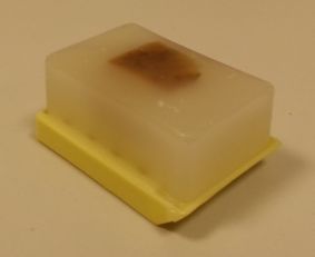
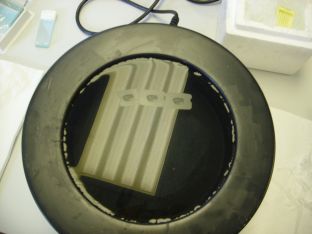
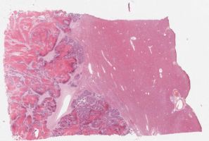
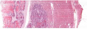
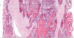
The process involves serially sectioning hundreds of sections onto glass slides from a piece of paraffin embedded tissue:
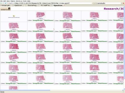
Then all these slides are stained and then scanned using Aperio software scanners creating virtual slides which are stacked and registered using specialised computer software:
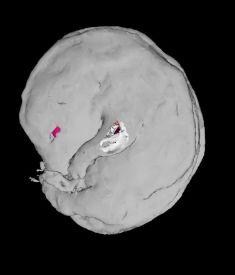
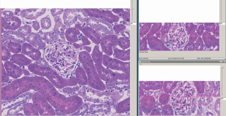
After registration these slices of liver with metastatic colorectal cancer can be viewed in the axial, coronal and sagittal planes. A large blood vessel near the tumour is highlighted in white:
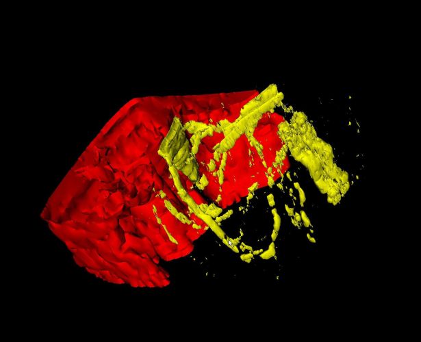
These stacks can then be visualised in 3-D which shows the carcinoma front in red and the blood vessels in the adjacent liver in yellow:
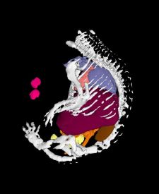
3-D reconstruction of whole sections is also possible, as demonstrated in this mouse embryo in which nearly 200 tissue sections were reconstructed to visualise the organs:
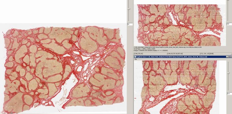
3-D reconstruction can also be used to visualise 3-D tissue models such as cirrhosis in the liver. This example shows Primary Biliary Cirrhosis (PBC) in the liver in which the cirrhotic nodules are stained pale yellow whilst the fibrotic scarring is stained red as a result of a Picro-Sirius red stain:
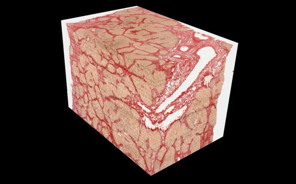
The model can be manipulated in the x, y and z axis in order for the pathologist to examine the cirrhosis in detail:
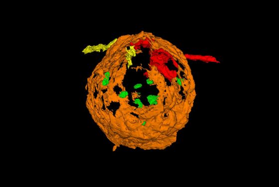
Because we are using high performance computing with virtual slides we can produce 3-D tissue reconstructions at a cellular resolution, such as this 3-D model of a single rat glomerulus:
The Bowman's capsule is reconstructed in 3-D (orange) with the proximal tubule exiting. The afferent (red) and efferent (yellow) arterioles can be seen exiting the capsule as well as the individual mesangial cells inside in green:
If you would like to know more or make use of our 3-D histopathology technologies please get in touch with Dr. Darren Treanor (darrentreanor@nhs.net)