10:15
|
Welcome, introduction, aims of meeting
|
|
10:20
| |
10:50
| |
11:20
| |
11:40
| |
12:10 - 13:00
Slides before lunch | | Case 1 | 61F. Jaundiced, very high ALT - 1700. Bilirubin 554. ? seronegative autoimmune hepatitis. |

Open with WebScope
| Case 2 | 62M. Jaundiced. ALT 2815, Bilirubin 560, AFP 11.6. Hepatitis A and CMV negative, other results awaited. |

Open with WebScope
| Case 3 | 21F. presented with acute liver failure, RUQ pain, ALT 1600, albumin 23. CT showed congestion, viral serology negative. On oral contraceptive for 4 years. Noted to have orogenital ulceration. Rapidly developed multi-organ failure, died on ITU. |

Open with WebScope
|
13:00
|
Lunch
|
14:00
|
Prof. W Rosenberg, Southampton
Non-invasive alternatives to liver biopsy |
|
14:30
| |
15:00
Slides before tea | | Case 4 | Referred at age 33 for injections sclerotherapy of oesophageal varices.
ERCP and liver biopsy consistent with sclerosing cholangitis affecting primarily intrahepatic bile ducts. No evidence of colitis. Started on Penicillamine.
Age 44 deterioration in liver enzymes. Hepatic angiogram revealed a 3 cm mass in upper part of the right lobe and an occluded portal vein. Alpha-fetoprotein raised to 700. Received intraarterial chemotherapy.
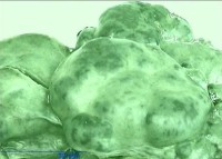
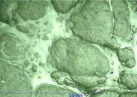
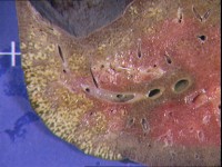
Liver transplantation a year later. Deeply bile stained cirrhotic liver with a subcapsular liver mass. Representative images of the mass and background liver. |

Open with WebScope
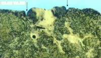
Macroscopy of liver bisected through the lesion (arrows indicate borders of the tumour).
Click image to enlarge
| Case 5 | Presented at age 3 weeks with diarrhoea + failure to thrive. Hospital admissions in infancy for respiratory problems. Paediatric Liver Unit referral for deteriorating liver function and ascites.
Biopsy showed marked steatosis + focal biliary type fibrosis.
Referral for end-stage liver disease.
Acute renal failure
Vancomycin-resistant Enterococcus sepsis
Malnutrition - Glucose intolerance
Refractory ascitis
Liver transplantation. |
|
15:30 |
Tea
|
15:50
| |
16:30
Slides before dinner
| | Case 6 | 48F. Philippino, lived in UK since age 16. Developed flu-like illness on holiday in Philippines, diagnosed pulmonary TB, started triple therapy (ethambutol, rifampicin, pyrazinamide) for 1 month then stopped.
One month later, back in UK, diagnosis of TB confirmed and commenced quadruple therapy (medication as above + isonizid). Two weeks later became jaundiced. Stopped drugs but developed coagulopathy, ALT 1100, increasing encephalopathy. Transplant 3 weeks after onset of jaundice. |

Open with WebScope
|
17:00 |
Close
|